

The urinary tract causes relatively infrequent issues in adult horses compared to other species (Schott et al, 2018). The following two-part article will discuss the approach to the more commonly encountered conditions and the treatment options available. Rather than an anatomical approach, conditions will be grouped by the predominant presenting clinical sign to optimise practical relevance. Part 1 will focus on abnormalities in the passage of urine including difficulty in passing urine (stranguria), increased frequency (pollakiuria) or volume (polyuria) of urination, and the inability to control the passage of urine (incontinence). There is considerable overlap between these signs and the differentiation of one from another in the clinical setting is not always easy. Part 2 will address abnormalities detected in the blood or urine itself, including azotaemia and pigmenturia. A detailed discussion of problems specifically associated with parturition is outside the scope of this article.
Stranguria
Stranguria describes difficulty in passing urine (Schott et al, 2018). In males, this may be recognised by repeatedly extruding the penis and posturing to urinate but with little or no urine voiding; females may repeatedly posture and evert the clitoris, again without passing a full urine stream. It is important to note that standing stretched out as if posturing to urinate occurs with abdominal discomfort; it is not uncommon for owners to describe this behaviour in horses with gastrointestinal forms of colic rather than stranguria. A complete history and physical examination are therefore prudent, especially in acute onset cases.
Stranguria, particularly in association with exercise, is a classic clinical sign of cystoliths. These are the most common form of equine uroliths (Laverty et al, 1992) and are primarily composed of calcium carbonate (Neumann et al, 1994). As well as stranguria, cases may present with pollakiuria, haematuria (especially after exercise) and urinary incontinence. Recurrent colic can also occur (Schott et al, 2018), as can weight loss (Laverty et al, 1992). Stones can be palpated per rectum or imaged using transrectal ultrasonography (Cercone, 2022). They are typically associated with small, empty bladders as a result of frequent urine voiding. However, if the bladder is full at the time of palpation, it is possible to miss the presence of a stone; therefore, emptying the bladder via urinary catheterisation may aid the diagnostic value of this technique. Blood analysis is expected to be normal, although anaemia, inflammation and azotaemia have been reported (Laverty et al, 1992). A detailed discussion of stone formation is beyond the scope of this article (Schott et al, 2018); however, damage to the uroepithelium is believed to be the most important factor (See and Williams, 1992).
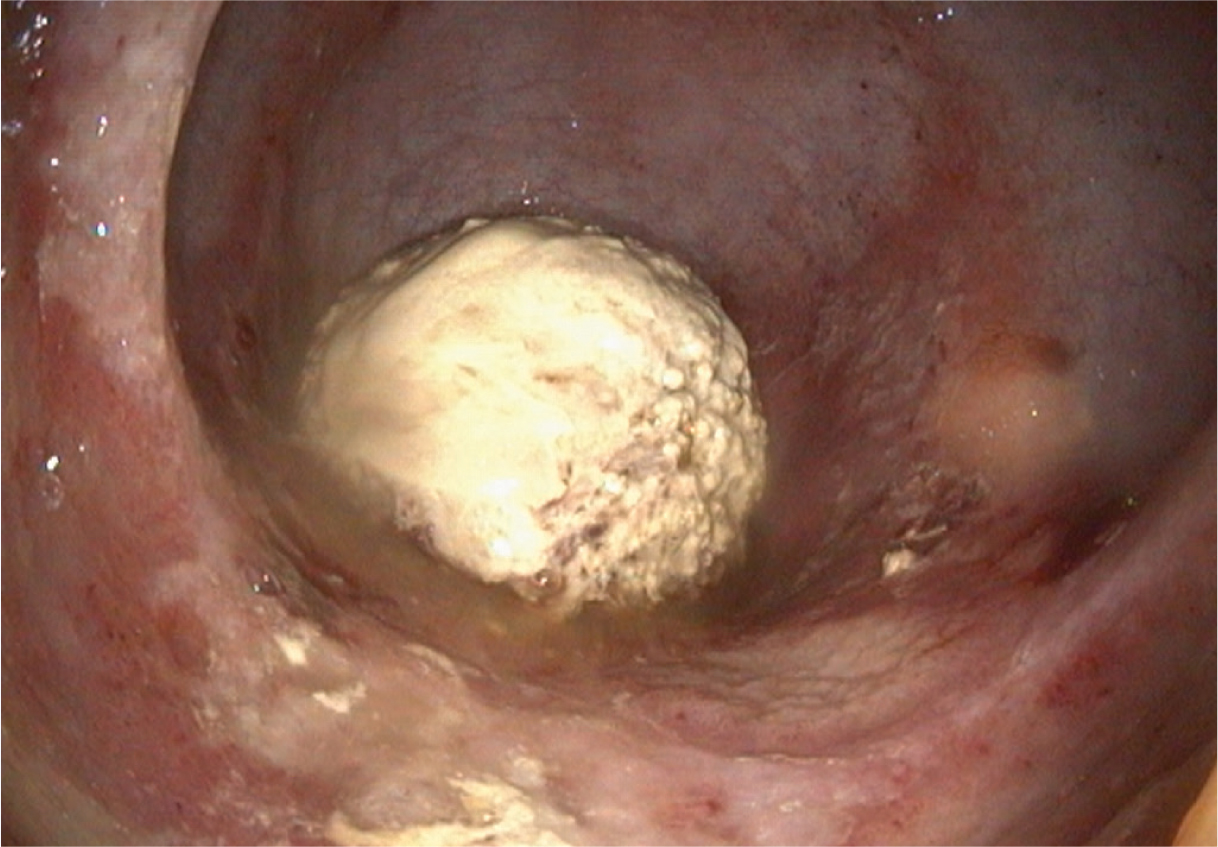
Cystoscopy is the mainstay of diagnosis (Box 1) as it enables direct visualisation of the size and number of calculi and assessment of the bladder mucosa (Cercone, 2022) (Figure 1). Stones are classified as type I and II; type I are usually ovoid, yellow-green in colour, spiculated and often easily broken up. Type II are less common, more irregular in shape but smooth-contoured (Schott et al, 2018). Stones may also be found in the ureters and/or kidneys. During cystoscopy, the ureteral openings should be assessed for size and symmetry, as abnormalities may suggest the presence of stones proximal to the bladder. Ultrasonography of the kidneys should also be performed to rule out nephrolithiasis and hydronephrosis. When distended, ureters may also be visible on ultrasound (Fubini and Delco, 2022). Assessing the renal parenchyma with ultrasonography immediately after cystoscopy can be hampered by presence of air from the bladder insufflation.
Cystoscopy
Equipment required:
Procedure:
Described complications:
Unlike in other species, dietary manipulation is not an integral part of management, and surgical removal is the mainstay of treatment. Various surgical options are available; which of these is performed will depend on the size of the calculus, sex and compliance of the patient, and surgeon preference. Laparocystotomy under general anaesthesia has been described via a caudal ventral midline (Lowe, 1961), caudal paramedian (Holt and Pearson, 1984) or parainguinal (Beard, 2004) approach. Laparoscopic cystotomy in dorsal recumbency (Ragle, 2000; Röcken et al, 2006) and a standing pararectal cystotomy approach are also described (Van Dongen and Plenderleith, 1994; Abuja et al, 2010).
Standing techniques via the urethra, under sedation and epidural anaesthesia, are commonly performed (Fubini and Delco, 2022). In mares, the short and distensible urethra allows for transurethral manipulation and removal, with or without crushing of the stones in situ. A longitudinal sphincterotomy may improve access (Fubini and Delco, 2022), though this approach is not possible in males. However, the creation of a temporary perineal urethrotomy allows the calculus to be grasped with forceps. Larger stones can be broken up before removal using various techniques including manual crushing (Frederick et al, 2012), fragmentation with a mallet and osteotome (Hawkins, 2013), pulsed-dye laser (Howard et al, 1998), holmium:yttrium-aluminum-garnet laser (Judy and Galuppo, 2002), electrohydraulic (Röcken et al, 2012) or ballistic shockwave (Koenig et al, 1999), and pneumatic impact lithotripsy (De Bernardis et al, 2019). Which option is selected will depend on surgeon preference and availability of equipment; the literature is limited to individual case reports or small case series, so a relative assessment of the various techniques is not available.
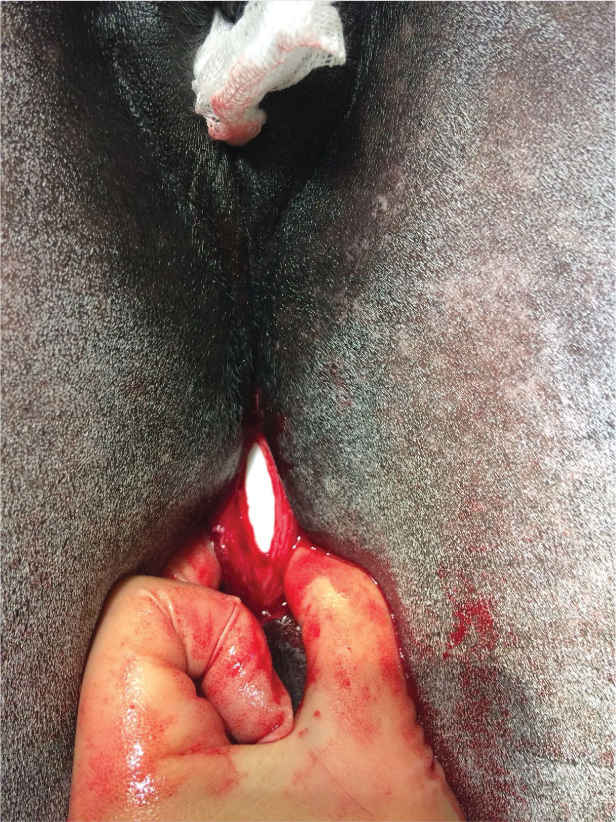
For larger stones, progressive urethral dilation may be beneficial (Sassot et al, 2023) and the use of a sterile bag (Figure 2) may minimise trauma to the urethral mucosa during extraction (Katzman et al, 2016; Williamson and McKinnon, 2017; De Bernardis et al, 2019). Following removal, the bladder should be copiously lavaged to remove debris that may act as a nidus for new stone formation. The urethral incision is typically allowed to heal by second intention; if performed, a sphincterotomy is closed routinely.
Complications reported include haemorrhage, persistent stranguria, formation of a stricture, a diverticulum or a fistula at urethrotomy sites, and recurrence of cystic calculi (Laverty et al, 1992; Kilcoyne and Dechant, 2014). The incidence of recurrence is debated; in the earlier literature, low rates of recurrence were reported (Lowe, 1965) but in later reports, clinical signs recurred in 41% of horses in a mean time of 13 months (Laverty et al, 1992). It may be that recurrence is more common following perineal urethrotomy rather than laparocystomy (Lowe, 1965; Laverty et al, 1992).
Following cystolith removal, anti-inflammatories and antimicrobials are typically indicated. The role of infection in the pathogenesis of equine cystoliths is unclear; a review of 68 cases found a positive urine bacterial culture in 10% of cases and a positive calculi culture in 63% (Laverty et al, 1992). Regardless, antimicrobials in the peri-operative period are typically recommended because of the trauma caused to the uroepithelium and the risk of translocation of bacteria into the bladder during the procedure.
Acidification of urine to <pH 6.5 reduces the propensity of calcium carbonate crystals forming and may reduce the risk of recurrence. Lowering the urinary pH can be achieved by lowering the dietary cation anion balance, for example by feeding ammonium chloride (DeBowes et al, 1984). However, results are not consistent, and even where pH is reduced, the reduction is often not sufficient enough to affect crystal formation (Nelson et al, 2020). Higher doses (up to 520 mg/kg per day of ammonium chloride) are more successful at lowering pH but are typically unpalatable and not suitable for long term use (Johnson and Crenshaw, 1990; Mair and Holt, 1994). High-grain diets typically have lower dietary cation anion balance and cause a lower urinary pH than forage-based diets, but the urine pH remains >7.0, so this is not a clinically relevant strategy (Wood et al, 1990). The amount of calcium in equine urine is proportional to the amount of calcium in the diet. As calculi are predominately calcium carbonate, it follows that avoiding feeding excessive amounts of calcium could reduce calciuria and may reduce stone formation (Schott et al, 2018). However, there is little evidence that this is an effective management strategy, and encouraging diuresis is likely to minimise stone formation. Daily salt feeding is often purported to do this; however, ponies fed up to 5% of total dry matter of the diet (approximately 375 g in a 500 kg horse) had no difference in water intake or urine production compared to controls (Schryver et al, 1987).
Calculi can also occur in the urethra. Where a complete obstruction has occurred, the horse will present with acute colic and achieving resolution is a medical emergency to prevent bladder rupture. If bladder rupture has already occurred at presentation, horses demonstrate signs of post-renal failure, including depression, anorexia and azotaemia (Trotter et al, 1981). Where partial obstruction occurs, the horse will present with stranguria.
Urethral calculi are less common than cystic calculi and are predominantly a male condition (Laverty et al, 1992). Stones typically lodge where the urethra narrows over the ischial arch or at sites of urethral stricture. Rhythmic contractions may be appreciable on palpation of the penis and the stone itself may be palpated (Trotter et al, 1981). Ultrasonography of the urethra will reveal an echogenic focus with acoustic shadowing consistent with the calculus itself (Cercone, 2022), and on palpation per rectum, a large, turgid bladder is felt. Passage of a urinary catheter is not possible, and the diagnosis is confirmed with endoscopy (Schott et al, 2018).
Where stones lodge in the distal urethra, gentle transurethral crushing with a hand or forceps is usually successful. However, for stones lodged at the ischial arch, a perineal urethrotomy is required (Schott et al, 2018). Fragmenting the stone in situ using radial extracorporeal shockwave therapy has also been successfully performed in two postmortem cases and one live animal (Verwilghen et al, 2008). Complications and post-operative management are as described for cystic calculi.
Bladder prolapse or extrusion through a tear in the vaginal floor can occur in mares, most commonly as a complication of foaling, and cause stranguria. In males, scrotal herniation of the bladder has been described in two foals, although pollakiuria was the main presenting sign (Cousty et al, 2010; Buyck et al, 2024). To the author's knowledge, this has not been described in adult horses. Stranguria has also been reported in one horse with an abdominal abscess and adhesions between the colon and bladder (Torske et al, 1992).
Pollakiuria
Pollakiuria describes an increase in the frequency of urination. This is distinct from polyuria, which refers to an increase in urine volume. It is also important not to confuse it with normal oestrous behaviour in mares. Any ailment that makes posturing to urinate difficult, such as back or hind limb pain, may result in pollakiuria as a result of aborted attempts to pass a full stream of urine (Schott et al, 2018).
Bacterial cystitis typically presents as pollakiuria. Stranguria, ‘urge incontinence’ and haematuria may also occur (Schott et al, 2018). Unlike with pyelonephritis, these horses will typically have normal haematology and biochemistry (Frye, 2006); ascending infection from the bladder to the upper urinary tract is uncommon but can occur with chronic, overdistension of the bladder.
The presence of inflammatory cells in the urine is suggestive, although not diagnostic, of cystitis. Gross examination of the urine is often unrewarding because of high levels of mucus and crystals in normal equine urine and wide variation in normal appearance (Schott et al, 2018). Leucocyte reagent pads on reagent strips (‘dipstick’) have not been validated for horses (Gratwick, 2021), so microscopic sediment analysis is required. An initial free catch sample containing increased white blood cells (>10 per high power field) and increased number of bacteria is highly suspicious of urinary tract infection. The sample should ideally be analysed rapidly (<60 minutes from collection) as bacterial numbers will increase at room temperature. To determine bacterial involvement, quantitative bacterial culture of the urine is required. To avoid contamination from the genitalia, a catheterised sample is typically preferred. Failing this, a mid-stream free catch should be used. A definitive diagnosis requires demonstration of >10 000 colony forming units per millilitre of urine. Isolation of multiple organisms is common, with typical isolates including Escherichia coli, Proteus spp., Klebsiella spp., Enterobacter spp., Streptococcus spp., Staphylococcus spp. and Pseudomonas aeruginosa (Schott et al, 2018). While the above confirms urogenital tract infection, it does not localise the site to the bladder. Pyelonephritis will also cause this but does not cause pollakiuria or stranguria; this will be discussed in Part 2.
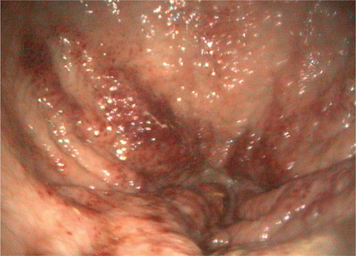
Primary cystitis is uncommon in horses; whenever cystitis is diagnosed, a predisposing/inciting cause should be suspected (Frye, 2006). Bacterial cystitis typically occurs as a result of conditions that alter urine flow and bladder emptying, including urolithiasis, bladder neoplasia, bladder paralysis or anatomical defects of the bladder or urethra. Iatrogenic causes are also possible, including urinary catheterisation or endoscopy, although should be suspected from the history (Schott et al, 2018). Further investigation is required to ascertain the cause. Palpation per rectum may identify uroliths or other bladder masses and transrectal ultrasonography enables assessment of the bladder wall. The ultrasonographic appearance of normal equine urine is echogenic and heterogenous; this should not be mistaken for sabulous material (Cercone, 2022). Ultimately, cystoscopy is likely to be the most useful diagnostic technique as it allows direct visualisation of the bladder mucosa, uroliths, neoplastic masses, sabulous material and other structures (Figure 3). Individual ureter samples can also be collected if there is any doubt regarding upper urinary tract infection.
Where possible, the first step in treatment of bacterial cystitis is to address the primary condition, such as by removing any cystoliths. Systemic antimicrobials are also required; these should be selected on the basis of culture and sensitivity, although empiric choices with urinary excretion include trimethoprim sulfadiazine, penicillin and aminoglycosides (Schott et al, 2018). Antimicrobials, such as trimethoprim sulfadiazine, are excreted by the kidneys and concentrated in the urine. This means that, although in vitro sensitivity may not suggest susceptibility, they may still be effective at the concentrations achieved in the bladder. In human medicine, instillation of the bladder with topical antibacterial agents such as oxychlorosene is reported (Messing and Stamey, 1978). To the author's knowledge, these have not been described in horses thus far but may be worth considering for refractory cases or where antimicrobial resistance is an issue. Phenazopyridine hydrochloride is widely used as a urinary tract analgesic in humans with urinary tract infections. Its use is described in horses (Aleman et al, 2011; Smith et al, 2018), although the safety and efficacy need further evaluation. In humans, acute kidney injury, methemoglobinaemia and haemolytic anaemia have all been described as complications (Messing and Stamey, 1978).
| Differential diagnosis | Key feature |
|---|---|
| Psychogenic polydipsia | Positive water deprivation test |
| Pituitary pars intermedia dysfunction | Increased basal adrenocorticotrophic hormone |
| Kidney disease | Azotaemia |
| Diabetes insipidus | Negative water deprivation test |
| Diabetes mellitus | Glucosuria |
| Sepsis | Clinical examination |
| Hepatic insufficiency | Biochemistry |
| Iatrogenic causes | Clinical history |
The duration of treatment is difficult to predict; weekly repetition of urinalysis for detection of pyuria and repeat urine culture is recommended. In experimentally-induced cystitis, resolution occurred within 6 days of starting trimethoprim sulfadiazine treatment or 2–4 weeks after bacterial inoculation in untreated horses (Divers et al, 1981). This study also highlights that, in the absence of an underlying cause, persistence of a bacterial infection is unlikely; where possible, correcting the predisposing condition, eg by cystolith removal, is key to management. In other species, the acidification of urine is considered beneficial for management of cystitis, but this is rarely achievable in horses.
Idiopathic haemorrhagic cystitis (Smith et al, 2018), polypoid cystitis (Rosales et al, 2019) and ulcerative cystitis secondary to phenylbutazone toxicity (Aleman et al, 2011) have been all described, but these present as haematuria, with pollakiuria only described in 1 out of 7 horses with idiopathic haemorrhagic cystitis and not with polypoid cystitis or ulcerative cystitis. This will be discussed in Part 2 of this series.
Polyuria polydipsia
When faced with the complaint of polyuria polydipsia, it is important to ensure polyuria is truly occurring. Polyuria is the passage of increased volume of urine and is typically defined as >25 litres of urine production per day (Schott et al, 2018). This is rarely actually measured in adults because of the technical challenges of urine collection. The description of an abnormally wet bed is one of the most useful indicators of polyuria and confirming polydipsia (increased drinking) by recording water intake exceeding 100 ml per kg body weight per day is practically achievable in most situations and is typically a good proxy for polyuria.
Blood analysis is a key starting point in the evaluation. Kidney disease can be identified by the combination of polyuria polydipsia with azotaemia. Where azotaemia is identified, a diagnosis of renal disease is likely; this will be covered in Part 2. In the older horse, blood analysis also allows identification of animals with pituitary pars intermedia dysfunction. Approximately one-third of pituitary pars intermedia dysfunction cases are believed to have polyuria polydipsia (Ireland and McGowan, 2018). A basal adrenocorticotrophic hormone test is the mainstay of diagnosis, although a thyrotropin-releasing hormone stimulation test can be performed where clinical suspicion remains (Stewart et al, 2023).
While sepsis is another cause of polyuria that would be expected to be accompanied by changes on blood analysis, this would not be the presenting or dominating clinical sign, nor would treatment be targeted at it, so it will not be discussed further. Hepatic disease has been described as a cause of polyuria polydipsia in the literature, but it is not routinely recognised (van Galen et al, 2024).
Iatrogenic causes of polyuria polydipsia, including alpha-2 agonists, corticosteroids, diuretics and intravenous fluid therapy, are possible but should be apparent from the history so will not be considered further. The hypothesised phenomenon of ‘medullary washout’ can result in polyuria following the cessation of intravenous fluid therapy. This has not been definitively confirmed in horses because medullary tonicity has not been measured, but it is recognised clinically (van Galen et al, 2024). Excessive salt consumption is described as a cause of polyuria polydipsia (Buntain and Coffman, 1981), although ponies fed large volumes of salt did not increase water consumption or urine output in one study (Schryver et al, 1987), so this is considered an unlikely cause except in the most extreme cases.
The most common cause of polyuria polydipsia is psychogenic polydipsia (Schott et al, 2018). The key aspect of this condition is that, although the horse produces hyposthenuric urine, it is capable of concentrating it. To assess this, a water deprivation test is performed (Box 2). In horses with long-standing psychogenic polydipsia, concentrating ability can be reduced because of presumed medullary washout. Therefore, a modified water deprivation test is required (Box 2) (McKenzie, 2007). An alternative to water deprivation testing is the Hickey-Hare test. This involves the infusion of hypertonic saline 2.5% (0.25 ml/kg/min for 45 minutes) to stimulate vasopressin release and water retention. A urine sample is then collected.
Water deprivation test
Aim:
Indications:
Procedure:
End points:
Modified water deprivation test:
For either a water deprivation test or a Hickey-Hare test, where the horse demonstrates the ability to concentrate its urine to >1.020, a diagnosis of psychogenic polydipsia is made. Management is aimed at reducing boredom and restricting water intake. Where the horse is unable to concentrate its urine, a diagnosis of diabetes insipidus is made. There are two forms of diabetes insipidus: neurogenic and nephrogenic. Although rare, both forms have been described in horses. Neurogenic diabetes insipidus is caused by the inability to produce or release anti-diuretic hormone (also known as arginine vasopressin) from the posterior pituitary. Described causes in horses include congenital (Kranenburg et al, 2010), encephalitis (Filar et al, 1971) and idiopathic (Breukink et al, 1983). Nephrogenic diabetes insipidus is caused by insensitivity of the renal collecting ducts to respond to anti-diuretic hormone for acquired (Morgan et al, 2012) or congenital reasons (Brashier, 2006), with an inherited, X-linked form suspected (Schott et al, 1993).
There are several methods to differentiate neurogenic and nephrogenic diabetes insipidus. Firstly, exogenous anti-diuretic hormone can be administered, either as an intravenous infusion (2.5 mU/kg over 60 minutes) or an intramuscular injection (0.5 U/kg). Alternatively, a synthetic analogue, desmopressin acetate, can be used at 0.05 μg/kg intravenously (Schott et al, 2018). The major limitation with both is availability; the author was unable to find an injectable source in the UK at the time of writing. The production of concentrated urine to >1.020 after drug administration is diagnostic for neurogenic diabetes insipidus; no change in urine concentration is seen with nephrogenic diabetes insipidus. Alternatively, endogenous anti-diuretic hormone can be measured at the conclusion of the water deprivation test, with high levels in nephrogenic diabetes insipidus and low/absent levels in neurogenic. To the author's knowledge, no commercial laboratory offers anti-diuretic hormone measurement.
Management of diabetes insipidus has most commonly been described as allowing constant access to unlimited water (Schott et al, 2018). However, for neurogenic diabetes insipidus, hormone replacement with desmopressin acetate has been successfully described in two cases; one in the short term using an eye drop formulation (Kranenburg et al, 2010) and one long term (>5 years) using daily subcutaneous injections (Durie and van Galen, 2020). Although described in humans (Nagae et al, 1994), recovery of anti-diuretic hormone secretion has not been described in horses, so management is likely to be lifelong, and the availability of desmopressin acetate may be a limiting factor. A nasal spray is commercially available, although to the author's knowledge, it has not been used in horses.
For nephrogenic diabetes insipidus, successful treatment has not been described in the literature. Extrapolating from other species, options to consider include thiazide diuretics, prostaglandin inhibitors and amiloride (Bockenhauer and Bichet, 2015).
Urinary incontinence
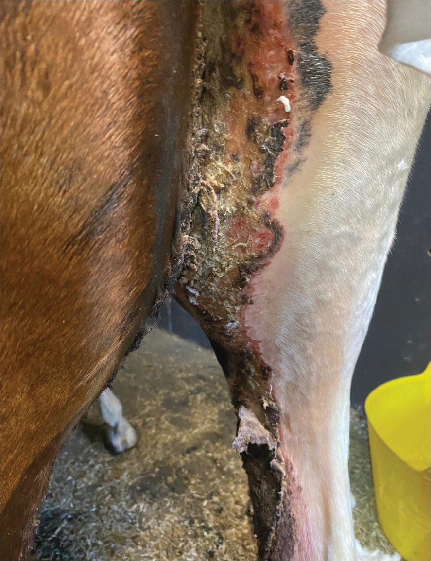
Urinary incontinence describes the involuntary passage of urine. It typically presents as constant or intermittent urine dribbling, often worsened by exercise or coughing, and urine scalding of the hindlimbs, perineum (female) (Figure 4) or ventrum (male) (Schott et al, 2018). Causes include intrinsic disease of the bladder and/or urethra and neurological disorders. A detailed description of the neurological control of continence and micturition is beyond the scope of this article and has been reviewed elsewhere (Mair, 2022). Congenital causes will not be discussed because of the focus on adult animals. However, it is worth noting that conditions such as ectopic ureters may not present until the horse begins exercise (Schott, 2011).
Neurological causes of urinary incontinence can involve the efferent or afferent nerves of the bladder and/or urethra, or may be intracranial in origin (Barrell, 2022). Therefore, a ‘whole horse’ approach is required and a full static and dynamic neurological examination should be performed in all cases.
In the UK, the primary infectious cause of neurological incontinence is equine herpesvirus-1 myeloencephalopathy. While uncommon (Aleman et al, 2018), it should always be considered because of the possibility of disease outbreak. Suggestive clues in the history include pyrexia and/or coughing in affected or in-contact horses, and the involvement of multiple horses. Urinary incontinence is extremely unlikely to be the sole neurological sign; ataxia typically predominates (Pusterla et al, 2022). Other infectious causes that are not endemic in the UK, but will occasionally present in horses with a history of foreign travel, include equine protozoal myeloencephalopathy. These horses will also typically have other clinical signs, such as ataxia (Aleman et al, 2018). Horses with ataxia as a result of equine degenerative myelopathy or weakness because of equine motor neuron disease may occasionally present with urinary incontinence as an additional clinical sign (Mair, 2022), although further discussion is beyond the scope of this article.
Polyneuritis equi (previously known as cauda equina neuritis) is a neurological cause of urinary incontinence in an otherwise apparently healthy horse (ie normal physical examination and blood analysis). The cause of this condition is unknown, although both autoimmune and viral causes have been suggested (Kadlubowski and Ingram, 1981). The initial clinical signs typically include perineal hyperaesthesia that presents as tail and perineal pruritus, and may be misdiagnosed as allergic dermatitis. Following this, hypalgesia/analgesia may occur, although will often be missed by the owner. Progression of disease results in paralysis of the tail, rectum, anus and bladder. Penile prolapse may be seen in males and muscle atrophy of the hindquarters may occur (Mair, 1990). This condition highlights the importance of a full neurological examination, as asymmetrical cranial nerve signs may occur and, when present, allow differentiation of this condition from more localised forms of urinary incontinence. Reported cranial nerve deficits include paralysis of the masticatory and facial muscles, head tilt, nystagmus, tongue paralysis and dysphagia (Mair, 2022).
Polyneuritis equi is currently a clinical diagnosis because there is no specific antemortem diagnostic test. Non-specific changes, such as xanthochromia and mildly increased cell count and total protein concentrations (Wright et al, 1987), may occur in the cerebrospinal fluid, and immunohistochemical abnormalities may be seen on muscle biopsy (Aleman et al, 2009). Definitive diagnosis requires post-mortem examination. There is currently no effective treatment (Mair, 2022). Corticosteroids may result in some temporary improvement in some cases, and bladder and rectum management are key if an acceptable quality of life is to be achieved.
In the absence of cranial nerve deficits, the main alternative neurological differentials for polyneuritis equi are sacral or coccygeal trauma, neoplasia of the lumbosacral spinal cord or idiopathic bladder paralysis (Mair, 2022). Lumbosacral intervertebral disc disease (Krueger et al, 2016) and vertebral osteomyelitis (Cudmore et al, 2012) have also been reported. History may be suggestive; for example, if the horse is known to have fallen or is a broodmare that has suffered dystocia, imaging of the caudal spinal column using radiography, scintigraphy or per rectum ultrasonography may be diagnostic.
When the lesion is cranial to the sacral segments of the spinal cord, upper motor neuron bladder dysfunction occurs. This is characterised by a taut, full bladder with high urethral resistance. However, because of the location of these nerves deep within the spinal cord, injury to them is generally associated with other catastrophic clinical signs, such as recumbency, that dominate the clinical picture and are often incompatible with life (Mair, 2022). Should treatment of upper motor neuron bladder dysfunction be undertaken, theoretical rationale exists for sympatholytic drugs such as phenoxybenzamine or prazosin to inhibit alpha-1 receptors in the smooth muscle of the internal sphincter (Labadia et al, 1988), parasympathomimetic drugs such as bethanechol to stimulate the detrusor muscle (King et al, 1998) and skeletal muscle relaxants such as dantrolene to reduce urethralis muscle tone (Mair, 2022). However, the clinical use of these drugs has not been critically evaluated in equine literature.
Lower motor neuron bladder dysfunction occurs when the lesion is in the sacral spinal cord segments, sacral spinal nerves, pelvic nerves, sacral plexus or pudendal nerves. This is characterised by a large, flaccid bladder that overflows, resulting in continual urine dribbling (Mair, 2022). This may be palpated per rectum and gentle pressure will easily express urine from it. There are typically other signs of dysfunction, such as a loss of external anal sphincter tone, reduced or absent perineal reflex, tail paralysis, perineal analgesia or hypalgesia, atrophy of the hind quarters and hind limb paresis. Occasionally, the penis or vulva also have a flaccid appearance (Mair, 2022). In such cases, the prognosis depends on the nature of the primary injury.
Where no cause is identified but the horse has urinary incontinence with a large, atonic bladder, a diagnosis of idiopathic bladder paralysis is made. Whether these truly represent an intrinsic failure in detrusor function or whether it is simply that the true inciting cause is not identified is unknown (Mair, 2022).
Various non-neurological causes of incontinence also occur. Damage to the urethral sphincter, most commonly following foaling trauma or as a result of iatrogenic causes, such as following removal of a cystolith, may result in urine dribbling. Diagnosis should be possible from the history and visual assessment, with or without endoscopy. Surgical repair may be possible in some cases (Schumacher and Brink, 2011). Oestrogen-responsive incontinence has been described in a handful of mares (Attenburrow and James, 1981; Watson et al, 1997). In other species, including dogs, oestrogen is believed to increase the sensitivity of urethral alpha-adrenergic receptors to increase internal sphincter tone (Kendall et al, 2024). In such cases, frequent administration of exogenous oestradiol appeared effective, although oestrogen preparations are not currently commercially available in the UK to the author's knowledge. Toxic causes of urinary incontinence have been described in horses grazing pastures containing species of sorghum and sudan grass (Knight, 1968; Van Kampen, 1970), but this has not been described in the UK.
Cystitis typically causes pollakiuria. However, it can result in urinary incontinence because of irritation of the bladder wall, resulting in stimulation of parasympathetic afferents and detrusor contraction (Mair, 2022). This is typically referred to as ‘urge incontinence’; converse to the incontinence discussed thus far, it is associated with detrusor overactivity. Alternatively, cystitis can occur secondary to other causes of incontinence such as idiopathic bladder paralysis or polyneuritis equi or their management.
Irrespective of the cause of bladder paralysis, sabulous urolithiasis is a common sequalae. Sabulous urolithiasis is the accumulation of sediment, primarily calcium carbonate crystals, in the bladder that can become inspissated. Over time, this accumulation of sediment causes chronic distension of the bladder and stretching of the detrusor muscle, reducing its ability to contract. Furthermore, the retention of urine leads to local ammonia production and mucosal irritation that predisposes to secondary cystitis. This leads to a perpetuating cycle of bladder dysfunction (Schott et al, 2018). It is this end-stage condition that the clinician is often presented with, precluding hopes of identifying the original cause or achieving a cure.
In short-term cases of bladder paralysis, eg those caused by equine herpesvirus myeloencephalopathy, the goal is to aid urine evacuation to avoid overdistension of the bladder. This maintains comfort and prevents detrusor muscle damage. This is best achieved with an indwelling urinary catheter. Prophylactic antimicrobials are typically considered because of the propensity to develop ascending bacterial cystitis.
Managing chronic cases requires significant owner commitment. Meticulous attention to perineal/abdominal and hind limb skin to prevent urine scalding is essential to maintain an acceptable quality of life. Minimising bladder distension to prevent further reduction in detrusor function is also important, although the use of a urinary catheter to achieve this is not practical because of the risk of ascending infection. Pharmacological treatments may be attempted, although their efficacy is questionable at best and by the chronic, end-stage situation that is often presented to the clinician, the effectiveness of any of these drugs is likely reduced (Mair, 2022). Bethanechol is a direct-acting parasympathomimetic drug that may aid detrusor function by stimulating muscarinic receptors (King et al, 1998). A wide dose range is described (0.2–0.4 mg/kg orally every 8–24 hours). Its bioavailability and efficacy with regards to the urinary tract has not been critically evaluated in horses, although improvement is anecdotally described at 0.04 mg/kg every 8 hours (Zakia et al, 2021).
Bladder lavage with isotonic crystalloids is likely necessary to remove accumulated sediment. This is typically performed in the standing horse, but has also been described under general anaesthesia. Passive infusion and pump-assisted delivery are reported (Rendle et al, 2008; Zakia et al, 2021). Lavage will require repeating because, unless the inciting cause is removed, sediment will continue to be deposited over time. The required frequency of this procedure is unknown; in a series of management of 13 cases, the median number of procedures per horse was two with a range of one to six. However, the period of time between lavages was not stated (Zakia et al, 2021). Repeated catheterisation has been associated with the development of a urethral stricture in one case (Rendle et al, 2008). The sediment may be firmly adherent to areas of bladder mucosal ulceration. Surgical removal via a urethrostomy or cystotomy is described, although not recommended (Schott et al, 2018).
Bacterial cystitis can occur secondary to sabulous cystitis or as a result of bladder lavage; therefore, systemic antimicrobials are typically administered. Although ascending infection from the bladder is uncommon in horses, bladder distension can cause incompetence of the bladder–ureter seal, resulting in uretitis and pyelonephritis (Fonteque et al, 2018). This will be discussed in Part 2.
The prognosis for urinary incontinence is generally considered poor, with many horses being euthanised as a result of problems associated with chronic/recurrent cystitis and/or ascending urinary tract infection. Management up to three years has been described in a handful of cases, although significant owner commitment is required (Rendle et al, 2008).
Conclusions
Abnormalities associated with the passage of urine are relatively uncommon in horses. When they do occur, they can be the result of urinary tract disease or disease in other body systems. A full physical examination and blood analysis should be performed as a starting point and in many cases, cystoscopy provides valuable diagnostic information. Prognosis varies from good with cystoliths to poor with bladder paralysis. Part 2 of this series will discuss abnormalities of the urinary tract associated with changes in the bloodwork or urine.