

In recent years, an increase in the emergence and re-emergence of vector-transmitted diseases has been seen across Northern Europe. Notable outbreaks include the arboviruses Bluetongue virus and Schmallenberg virus, both of which affected the UK ruminant population in the winter of 2023–2024, generating significant financial and welfare implications (Gray, 2024; Jusuf et al, 2024). Though endemic to sub-Saharan Africa, African Horse Sickness virus also presents an increasing threat to the UK equine population, with outbreaks reported in Northern Africa, South-East Asia and Europe in recent decades (Carpenter et al, 2017). These viruses are all non-contagious and transmitted by Culicoides midges. Research indicates an increasing abundance of these important vectors in the UK, with a lengthening of the Culicoides adult activity season (Sanders et al, 2019) and over-wintering of larvae recently reported (Stokes et al, 2022). Another Culicoides midge-transmitted pathogen which may present an increasing threat to the UK equine population is the filarial nematode Onchocerca cervicalis.
Onchocerca cervicalis (also referred to as equine neck thread-worm) is the causative agent of equine onchocerciasis, which commonly manifests as a dermatitis characterised by severe pruritis and subsequent skin lesions, thus presenting significant welfare implications. Diagnosis of equine onchocerciasis is complicated by a lack of effective diagnostic assays and the fact that the condition is a differential diagnosis for several common equine skin conditions, including insect bite hypersensitivity. There is lack of research and discussion of O. cervicalis in British literature, and survey work has suggested that practising equine vets may lack knowledge of the nematode's lifecycle (Mansell and Behnke, 2022), highlighting the need to increase knowledge and consideration of this important parasite.
Onchocerca cervicialis lifecycle and epidemiology
O. cervicalis is a filarial nematode with an indirect lifecycle (Figure 1). Culicoides midges act as the dominant intermediate host, and Culicoides nubeculosus has been identified as the major vector in the UK (Mellor, 1975). O. cervicalis has worldwide distribution, affecting horses, donkeys and mules across the globe (Ottley et al, 1983; Collobert et al, 1995; Marques and Scroferneker, 2004; Papini et al, 2020). The parasite has been most widely documented in the United States, where Culicoides variipennis has been identified as a major vector (Foil et al, 1987). In tropical regions, mosquitoes and Simulium (black flies) have also been identified as potential intermediate hosts (Lok et al, 1980; Marques and Scroferneker, 2004).
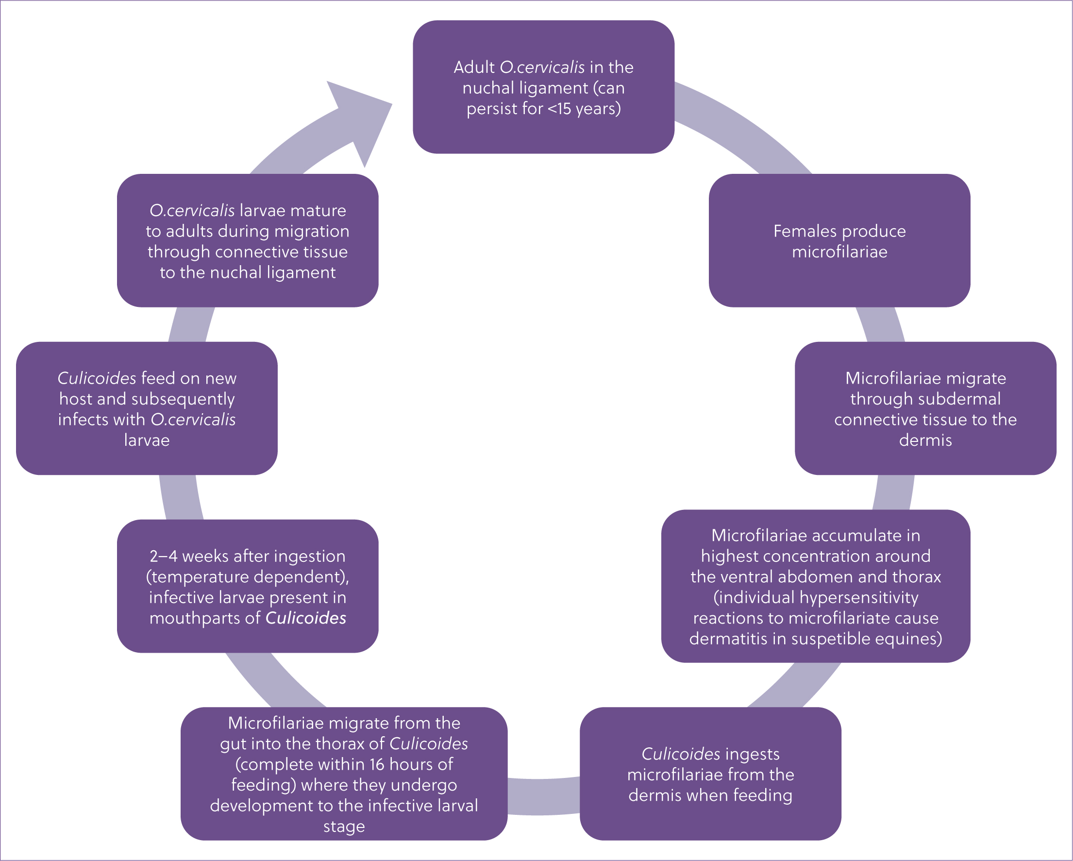
Adult O. cervicalis reside in the funicular region of the nuchal ligament (often between the last cervical and forth thoracic vertebrae), where they may persist for up to 15 years. The females are long (30–50 cm), thread-like and usually coiled up within nodules. They are viviparous and produce first stage larvae, or microfilariae, which measure approximately 250 µm in length. The microfilariae migrate through connective tissue to the dermis, accumulating in highest concentrations around the ventral abdomen and thorax (Mellor, 1973a). This corresponds with the preferential landing and biting sites of Culicoides and is considered to be an evolutionary adaptation of the parasite to accommodate the vectors feeding habits (Mellor, 1974). Microfilariae may accumulate in smaller concentrations around the face, neck and withers.
Recent epidemiological research into the prevalence of O. cervicalis globally is lacking, and reported infection rates vary considerably between studies depending on a number of factors including geographical location, diagnostic method and the age of animals sampled. Older equines are more susceptible to O. cervicalis infection; therefore, abattoir studies involving examination of the nuchal liga ment likely report higher infection rates than would be found in the general equine population (Schmidt et al, 1982; Lyons et al, 2000). The last notable research on O. cervicalis in the UK was conducted 50 years ago (Mellor, 1973a), where post-mortem examination of nuchal ligaments alongside skin samples found infection rates of 22.7% and 13.9% at two abattoirs in the South East of England. An infection rate of 12.9% was found at a third abattoir based on the presence of microfilariae in skin samples only.
Equine onchocerciasis
Clinical signs
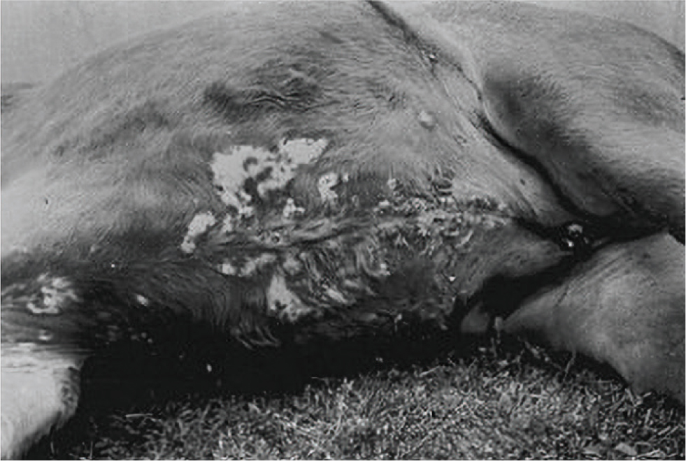
Equine onchocerciasis is the clinical syndrome caused by microfilariae infection. Adult O. cervicalis were historically associated with the development of fistulous withers (associated with nuchal ligament inflammation), but are now considered to have minimal or no pathogenic effects. Equine onchocerciasis most commonly manifests as dermatitis, characterised by severe pruritis and subsequent skin lesions in the areas where dermal microfilariae congregate (notably the ventral abdomen and thorax; Figure 2). These clinical signs are hypothesised to result from individual type I and type III hypersensitivity reactions to dermal microfilariae antigens, and eosinophil infiltration is often found alongside microfilariae in skin samples. The gross appearance of lesions varies between case studies, with areas of alopecia, scaling, crusting and leukoderma (depigmentation) often seen (Rashmir-Raven, 2018). As the condition progresses, ulceration and lichenification may occur and secondary bacterial infections can develop (Nielsen and Reinemeyer, 2018). As such, equine onchocerciasis can present significant welfare implications, with pruritis in particular known to impair quality of life and the ability to express normal behaviours (Marsella et al, 2023).
Treatment and management
No treatments are effective at eliminating adult O. cervicalis, which can persist in the nuchal ligament for several years. The macrocyclic lactones ivermectin and moxidectin are highly effective at killing dermal microfilariae and supressing microfilariae production by adults, so regular administration of these products can control the clinical signs of equine onchocerciasis. Initially (<72 hours post treatment), clinical signs may be exacerbated and/or ventral oedema may develop, particularly following ivermectin use. These seemingly adverse reactions are thought to be because of dead or dying microfilariae antigens initiating a greater inflammatory response (Nielsen and Reinemeyer, 2018). However, regression of lesions and absence of microfilariae from skin samples are usually observed within 1–2 weeks of treatment (Pollitt et al, 1986; Mancebo et al, 1997).
Diagnosis
Diagnosis of equine skin conditions is often challenging as clinical signs greatly overlap. Equine onchocerciasis is no exception and acts as a differential diagnosis for several common skin disorders, including insect bite hypersensitivity (Marsella et al, 2023). The two conditions may occur simultaneously, with insect bite hypersensitivity resulting from type I and type IV hypersensitivity reactions to Culicoides midge saliva (Cox and Stewart, 2023). The seasonal nature of insect bite hypersensitivity may enable differentiation to an extent, as the clinical signs of equine onchocerciasis generally occur throughout the year. However, equine onchocerciasis lesions can increase in severity during Culicoides activity season (Nielsen and Reinemeyer, 2018). Depending on the Culicoides species involved, insect bite hypersensitivity lesions are usually most prevalent around the mane, crest and tail with the ventral abdomen less commonly affected (Cox and Stewart, 2023). In some case reports, equine onchocerciasis lesions have also been concentrated in the neck and tail-head regions (Solcan et al, 2018). In the absence of appropriate diagnostic tests, response to treatment is likely the most common method of differentiation between insect bite hypersensitivity and equine onchocerciasis. Macrocyclic lactone administration will fail to resolve, or exacerbate and subsequently resolve, the clinical signs of insect bite hypersensitivity, while standard treatment and management practices for insect bite hypersensitivity (systemic or topical corticosteroids, systemic antihistamines and Culicoides avoidance) will be ineffective for equine onchocerciasis. Other likely differential diagnosis for equine onchocerciasis, which may also occur simultaneously, include lice infestations, contact dermatitis and pyodermas.
The only ante-mortem test available for equine onchocerciasis is microscopic examination of skin samples to detect dermal microfilariae. The sensitivity of this method is highly variable (Bottomley et al, 2016) depending on the number and size of samples taken, microfilariae distribution and the time since ivermectin or moxidectin was last administered. Furthermore, such procedures can be costly, time-consuming and perceived as invasive by clients.
Diagnosis is further complicated by the fact that equines are often infected with O. cervicalis asymptomatically (Mellor, 1973b) and as such, microfilariae detection in an individual suffering from skin disease does not guarantee that O. cervicalis is the causative agent. In recent years, a finger-prick lateral-flow assay to detect antibodies against Onchocerca volvulus (the causative agent of river blindness in humans) has been made available with 89.1% sensitivity (Golden et al, 2013; Vlaminck et al, 2015). Along with the recent developments made in Anoplocephala (tapeworm) and cyathostomins (small redworm) diagnosis for equines (Matthews et al, 2023), this presents opportunities to develop highly sensitive, non-invasive and cost-effective diagnostic tests for O. cervicalis in the future.
The UK situation
The last notable study on O. cervicalis in British horses was published over 50 years ago at the time of writing, finding infection rates of 22.7%, 13.9% and 12.9% across three abattoirs (Mellor, 1973a); however, the authors noted that none of the infected horses exhibited skin lesions which could be definitely attributed to the presence of dermal microfilariae (Mellor, 1973b). This study was conducted before macrocyclic lactones became available in the veterinary market, and Culicoides activity in the UK has changed since in response to increased temperatures and precipitation (Sanders et al, 2019). Therefore, the current prevalence of O. cervicalis in the UK equine population is unknown.
Since becoming licensed for use in the UK, macrocyclic lactones have become the most common anthelmintic products administered to UK horses. A sample of 463 UK horse owners and yard managers reported that either ivermectin or moxidectin products had been used on their premises in the previous 12 months (77.5% and 67.6% respectively; Tzelos et al, 2019). Current best practice guidelines (Rendle et al, 2019) state that moxidectin should be administered as part of the quarantine procedure when adult horses are moved onto new premises, and should also be administered to horses classed as moderate or high risk for cyathostomins in mid–late autumn to remove both adult and larval stages. The guidelines also state that ivermectin can be administered throughout the grazing season in response to high faecal egg counts. Considering that moxidectin and ivermectin are routinely administered to UK horses, it is likely that equine onchocerciasis is being inadvertently controlled and therefore is presumed rare and of minor importance in the UK.
In the winter of 2019-2020, an online survey was conducted with the aim of assessing the current awareness and knowledge of O. cervicalis amongst UK equine vets (Mansell and Behnke, 2022). The survey had a low response rate, with only 88 responses obtained and analysed; this limits the external validity of the findings and places limitations on the conclusions that can be drawn. However, the study did provide valuable insight and highlight the requirement for further research in this area. The key findings from the survey are shown in Figure 3. While the majority of respondents were aware of O. cervicalis, the results of the survey highlighted the need to increase knowledge and consideration of the parasite as a potential cause of skin disease. The lack of recognition of the clinical signs of equine onchocerciasis likely explains why most respondents did not consider it as a differential diagnosis. The high prevalence of ‘unresponsive’ insect bite hypersensitivity reported was a cause for concern; a proportion of these cases could be undiagnosed equine onchocerciasis, although further research is required to confirm this hypothesis. When investigating cases of unresponsive or ‘difficult to manage’ insect bite hypersensitivity, it is important that anthelmintic treatment history is considered.

The survey findings also present a number of implications for the wider equine veterinary industry. These include the requirement to develop effective, practical assays for use in the diagnosis of equine onchocerciasis and wider surveillance of O. cervicalis. Such tests would inform targeted use of ivermectin and moxidectin and help to preserve the e^cacy of these products, while also enabling differentiation of equine onchocerciasis from unresponsive or ‘di^cult to manage’ insect bite hypersensitivity.
What's next?
Equine parasite control guidelines in the UK are currently being reviewed. Considering increasing reports of resistance to moxidectin in equine nematodes, including in cyathostomins (Bull et al, 2023), alongside the recent launch of a serum antibody test for cyathostomins (Matthews et al, 2023), it is likely that treatment recommendations will be altered towards more targeted use of the anthelmintic. Similarly, ivermectin use will likely decline further in the near future as a result of increasing uptake of faecal egg counts and non-chemical parasite control strategies by horse owners (Tzelos et al, 2019). There is also the risk of O. cervicalis itself developing resistance to the macrocyclic lactones, with reduced ivermectin e^cacy against O. volvulus recently observed in humans (Nielsen, 2022). If this happens and Culicoides midge abundance continues to increase, then the number of equine onchocerciasis cases in UK horses could rise considerably in the coming years. As such, there is a need to increase awareness and knowledge of O. cervicalis within the veterinary profession, while also conducting further research to determine the parasites current prevalence in the UK and improve diagnosis.
Conclusions
Onchocerca cervicalis may become more problematic in the UK equine population in the near future, especially considering increasing Culicoides abundance and declining prophylactic use of macro-cyclic lactones. There is a lack of research on O. cervicalis in the UK and diagnosis is challenging, with equine onchocerciasis acting as a differential diagnosis for several common equine skin conditions. Findings from recent survey work suggest that UK-based equine vets often do not consider O. cervicalis as a potential cause of skin disease, highlighting the need to increase awareness and knowledge of O. cervicalis within the veterinary profession.